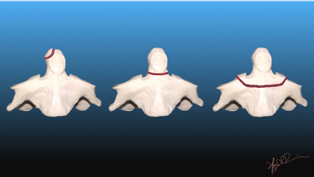

Masses this would be most concerning for a Jefferson (or Burst) fracture of C1. If the lateral masses of C1 extend out beyond the C2 lateral Congenital C1 arch absence, however, is a very. Spaces between the dens and the lateral masses of C1 Abstract: Odontoid fractures are one of the most common injuries to the cervical spine in geriatric patients. Spaces between the lateral masses of C1 and the body of C2 (axis). Fractures of the odontoid peg of the cervical spine Introduction Management of odontoid fractures has been recognised as a challenge since the first description of these injuries in the early 20th century 1. Make sure there is no asymmetry of the articular Furthermore, an MRI is preferred over a CT scan, since the CT scan may not be able to show the maximal positions of displacement in the fractures. We review the case of a 92-years-old man with traumatic Grauer type II B odontoid fracture treated with anterior cannulated screw fixation. There are varied management approaches with paucity of robust evidence to guide decision-making. Diagnosis can be made with standard lateral and open-mouth odontoid radiographs. Odontoid fractures constitute the commonest cervical spinal fracture in the elderly. However, mortality rates of older individuals with odontoid or subaxial spine fractures. Odontoid Fractures are relatively common fractures of the C2 (axis) dens that can be seen in low energy falls in elderly patients and high energy traumatic injuries in younger patients. While this radiographic rule can be used, it is important to recognize that it may not always correlate well and management decisions should not be made without first obtaining an MRI. Cervical spine fractures can lead to many devastating consequences. The rule of Spence would suggest that if there is more than a combined (total of both sides) overhang of 6.9 mm or more of the lateral masses of C1 in relation to the C2 lateral masses then there is concern for an injury to the transverse ligament and an MRI should be done. The reactive new bone formation around the odontoid fracture may play a role in preventing further movement and development of myelopathy.Make sure the lateral masses of C1 (atlas) do not hang over the lateral masses of C2 (axis).Note: Scroll over or tap over image to see lines & labels.


 0 kommentar(er)
